Overview
In an estimated 20 to 30% of persons with hepatitis C virus (HCV) infection, persistent liver inflammation leads to fibrosis and eventual development of cirrhosis.[1,2,3] Advanced fibrosis and cirrhosis—at their early stages—are not usually clinically apparent or symptomatic.[4] As individuals develop more extensive hepatic fibrosis, physiologic complications can develop, such as increased pressure within the portal system, disruption in bilirubin metabolism, and reduced production of coagulation factors and other proteins.[5,6] This lesson discusses the clinical considerations that come with the care of a person with HCV-related cirrhosis.
Defining Compensated and Decompensated Cirrhosis
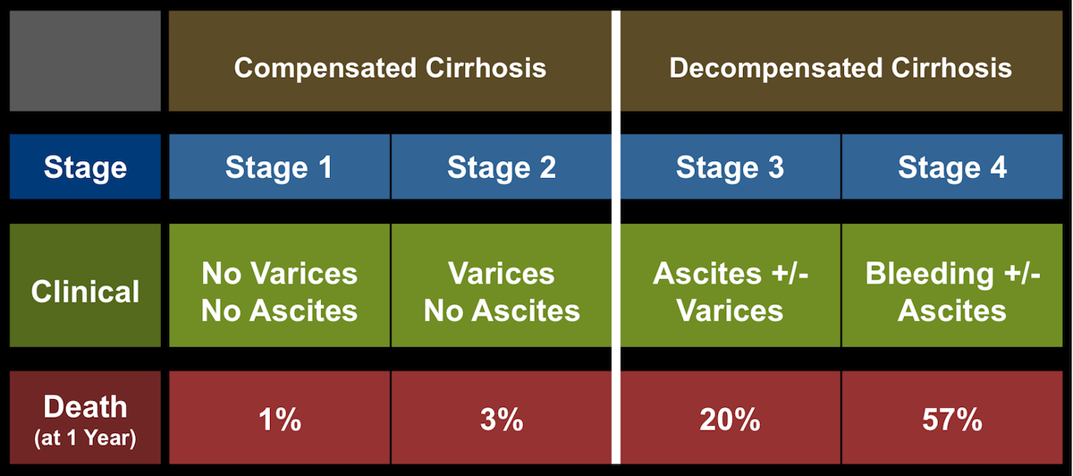
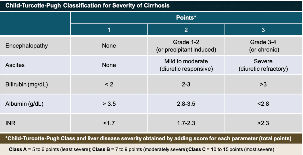
Once it has been established that an individual has cirrhosis, it is very important to determine whether they have compensated or decompensated cirrhosis.[7] Persons with compensated cirrhosis often do not have signs or symptoms related to their cirrhosis, although they may have evidence of portal hypertension, such as esophageal or gastric varices.[8,9,10] In contrast, persons with decompensated cirrhosis often have symptomatic complications related to cirrhosis, including those related to hepatic insufficiency (jaundice or hepatic encephalopathy) and those related to portal hypertension (ascites or variceal hemorrhage).[11] Some experts have proposed a 4-stage cirrhosis classification system to risk-stratify individuals according to the presence of ascites, esophageal varices, and variceal bleeding to differentiate and stage compensated and decompensated disease, although the Child-Turcotte-Pugh score (discussed later in this lesson) is more widely used (Figure 1).[12,13]
Importance of Distinguishing Compensated versus Decompensated Cirrhosis
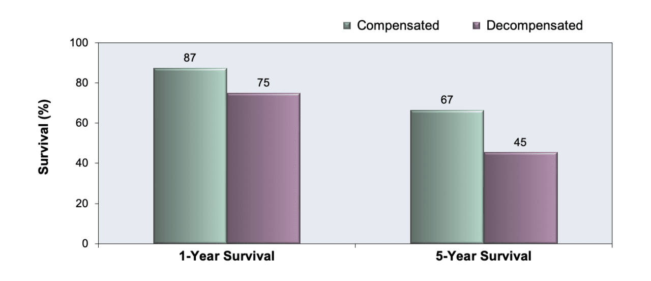
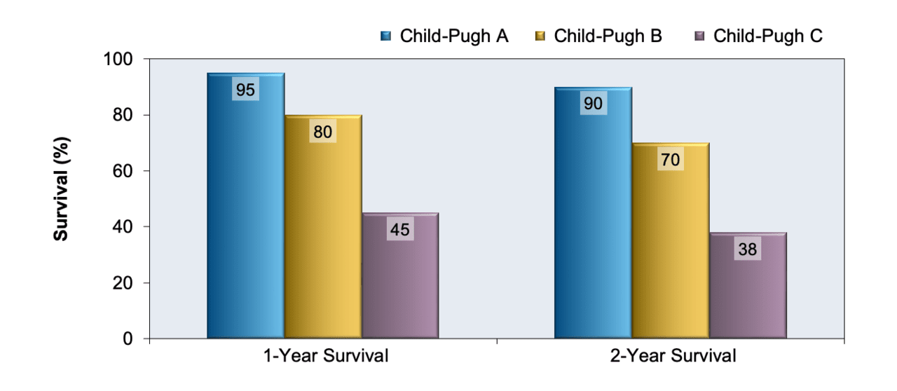
Prognosis and survival are markedly better in persons with compensated cirrhosis than in those with decompensated cirrhosis (Figure 2).[14,15] In addition, the presence of decompensated cirrhosis can have major implications regarding management and prevention of cirrhosis-related complications, as well as the potential need for a referral for liver transplantation evaluation.[16] In general, any person with decompensated cirrhosis should receive evaluation and medical care from a hepatologist or liver diseases specialist.[7]